Micrograph Gallery
Here are some of the beautiful micrographs captured by our RBSM members
-
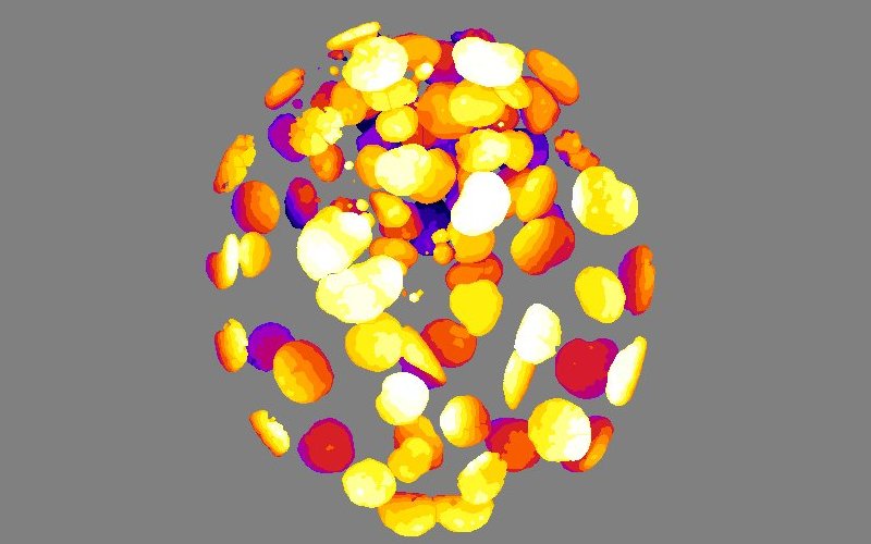
Embryo - DAPI counterstained nuclei of a human embryo, segmented and depth-color-coded.
© Winnok De Vos, University of Antwerp -
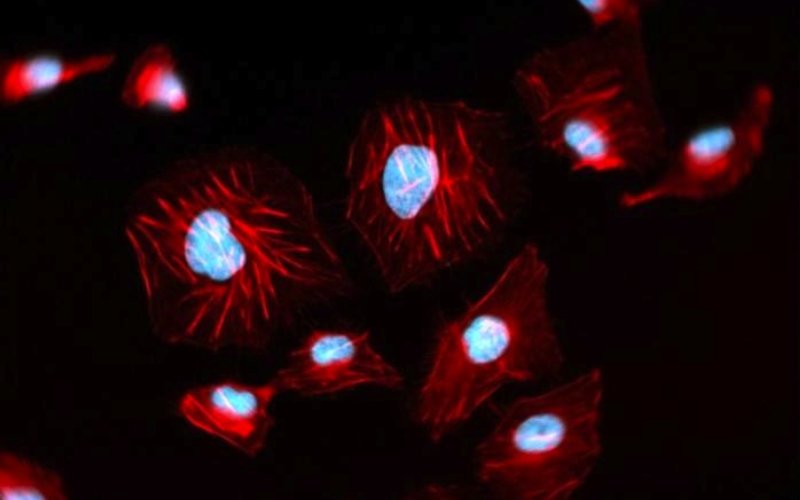
Actin cytoskeleton - Widefield Microscopy of phalloidin/DAPI counterstained ECV-304 cells
© Winnok De Vos, Antwerp University -
Harmonic - Mitotic cells visualised by autofluorescence (red) and second harmonics (green).
© Marcel Ameloot, Biophysics group, Hasselt University. -
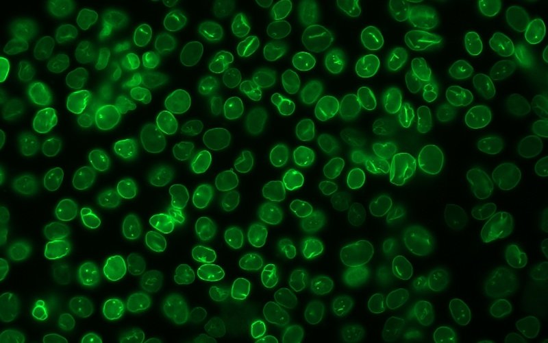
ECV-LMNA - Widefield fluorescence image of stable transgenic ECV-LMNA cells
© Winnok De Vos, Antwerp University -

Root tip - Confocal reconstruction of transgenic Arabidopsis thaliana H2B-GFP plant.
© Winnok De Vos, Antwerp University -
BY2 cells - Confocal section of BY2 tobacco cells transformed with a GFP-Nictaba construct.
© Annelies Delporte, Ghent University -
Nuclear dysmorphy - Compound progeroid patient cells counterstained for nuclear envelope components lamin B1 and NPC's.
© Winnok De Vos, Antwerp University -
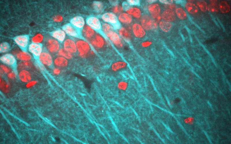
Hippocampus - Confocal section of cleared Thy1-GFP (cyan) mouse brain counterstained for neuronal nuclei (red)
© Jan Detrez, Antwerp University -
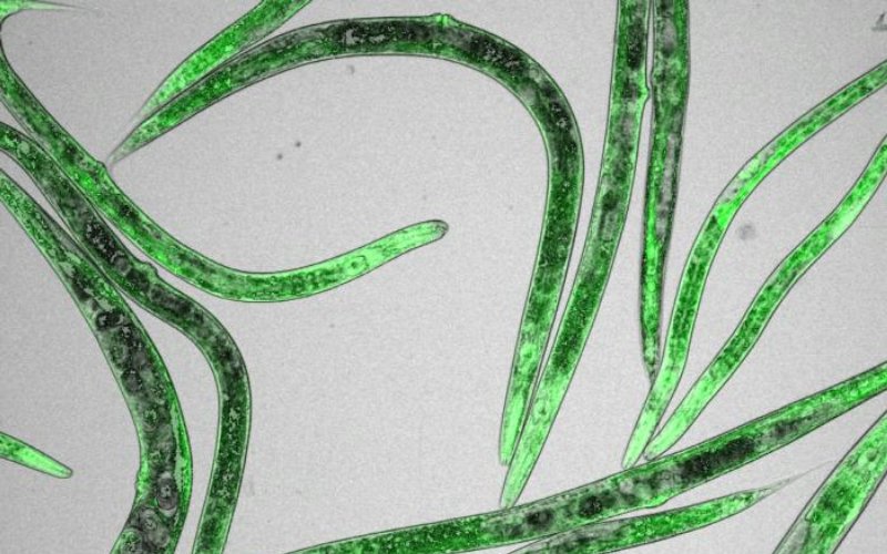
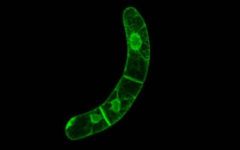
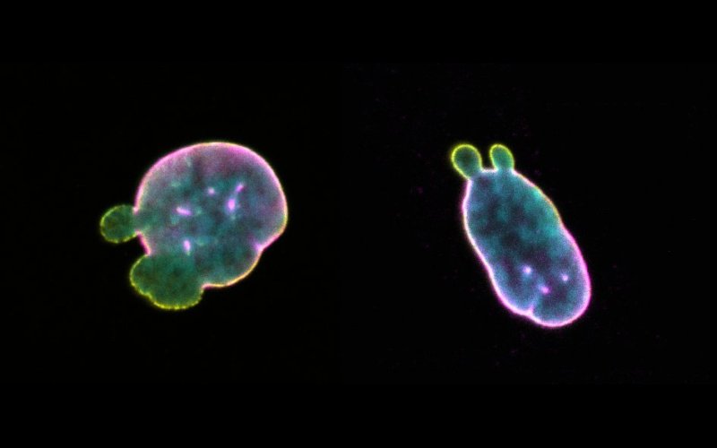
Hyper worms - Combined transmitted and fluorescence image of a C. elegans strain expressing HyPer-GFP
© Patricia Back, Ghent University -
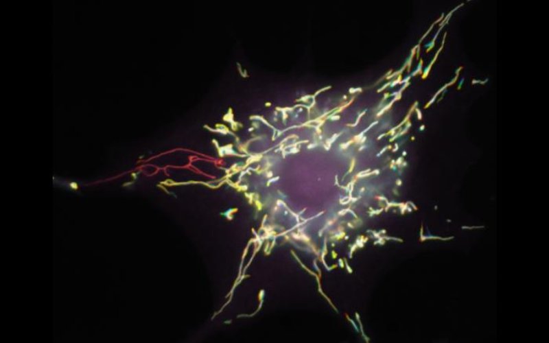
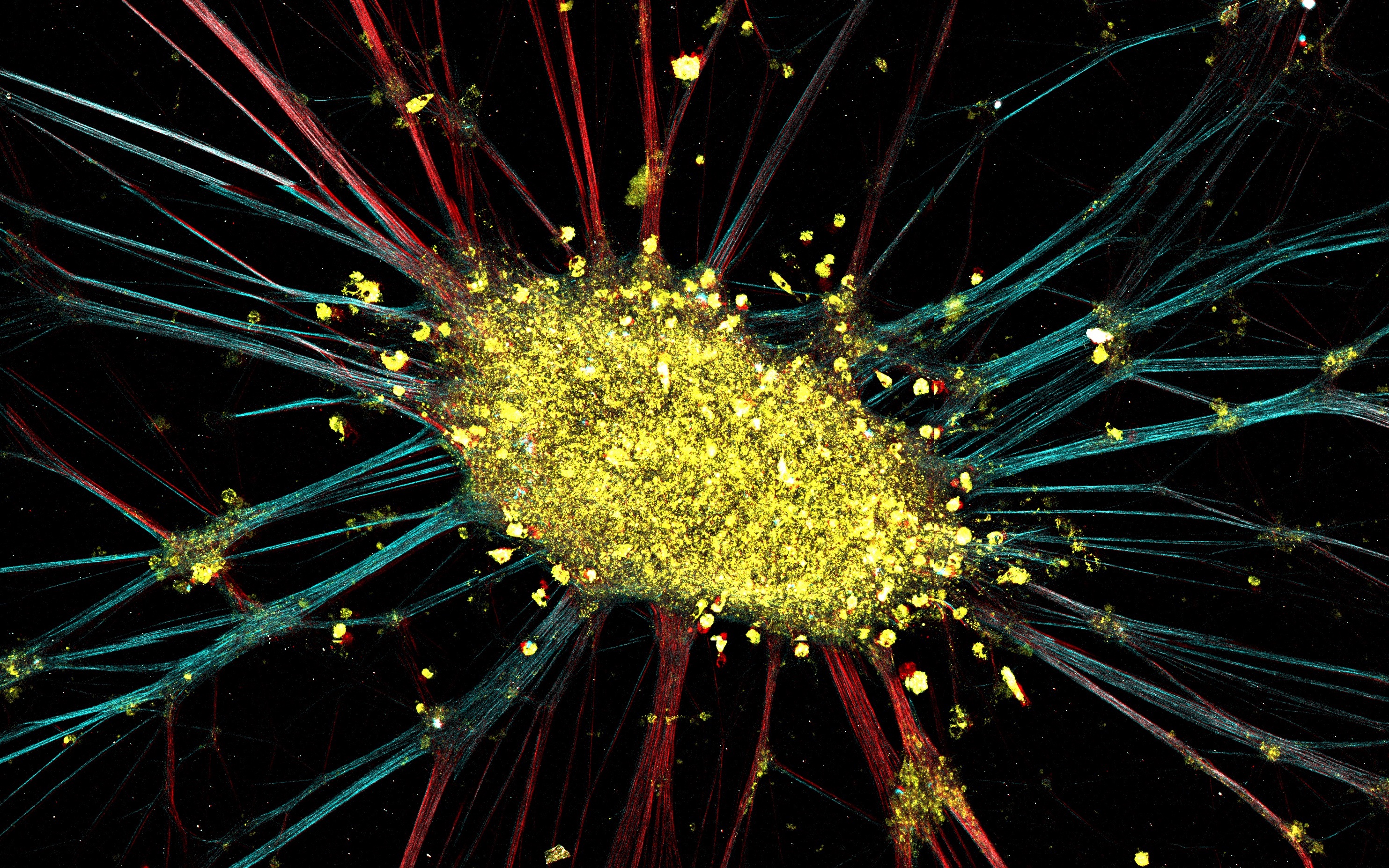
Short circuit - Color-coded time projection of a human fibroblast undergoing mitochondrial depolarization.
© Tom Sieprath, Ghent University -
Cross - DNA damage repair in a U2OS-53BP1 cell nucleus after laser micro-irradiation.
© Winnok De Vos, Antwerp University. -
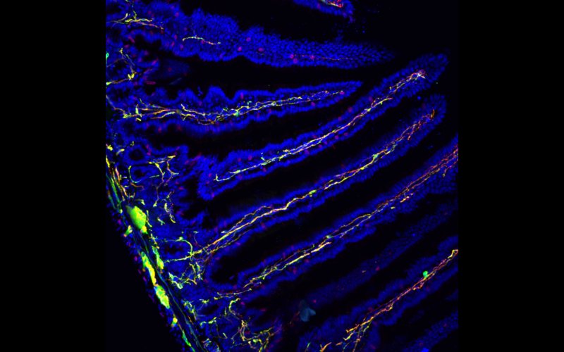
Innervation - Mouse intestine section stained for enteric neurons/glia (green), neuronal processes (red) and nuclei (blue).
© Zhi-ling Li, KUL (RBSM awardee 2016) -
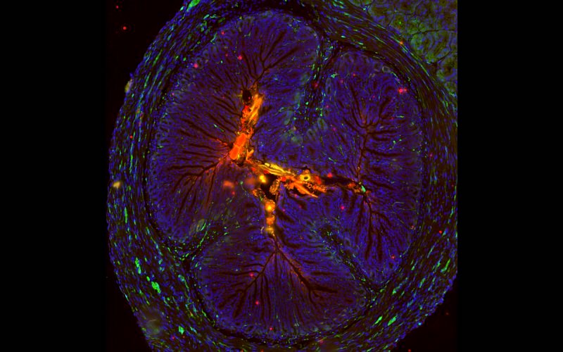
Insect look-a-like - Spry4 KO mouse antrum section stained for S100β+ glia (green), enteric neurons (red) and nuclei (blue).
© Pierre Vandenberghe, ULB (RBSM awardee 2016) -
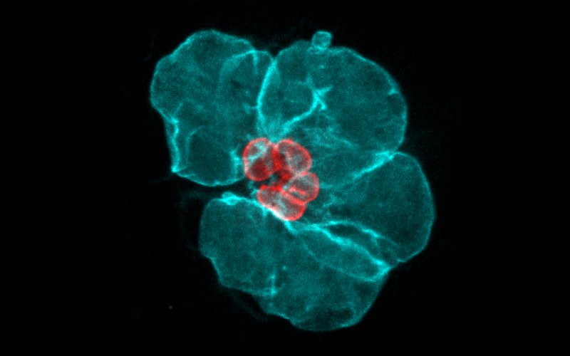
Expanded Nuclei - Overlay of lamin-counterstained carcinoma nuclei before (red) and after (cyan) expansion.
© Joke Robijns, Antwerp University (RBSM awardee 2018)
Spotlight: RBSM 2018 Picture Awards
The more the merrier -
Label-free image of neuroblastoma formation. Polarization-dependent forward SHG reveals horizontally (cyan) or vertically oriented (red) microtubule bundles in axonal projections. Autofluorescence (yellow) from the cell coma was acquired simultaneously.
© Valérie Van Steenberghen, KU Leuven
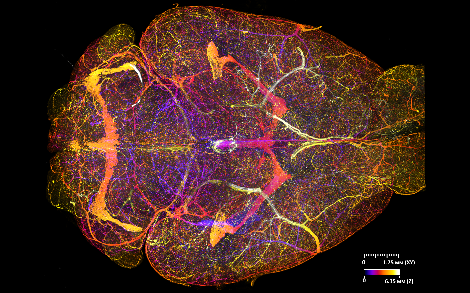
Innervated brain -
Light-sheet microscopy of optically cleared mouse brain perfusion stained for the vasculature. Fluorescent labelling was done by intravenous administration of a Alexa647-conjugated lectin. A colour-weighted axial projection was rendered with a Fire lookup-table to visualize depth differences.
© Jan Detrez, UAntwerpen
Want to participate?
Send in your microphotographic art and win the RBSM picture award...